
Case of the Month June 2021
A 77-year-old man taking the proton pump inhibitor (PPI) pantoprazole for several years to treat gastroesophageal reflux underwent an upper endoscopy during which a polyp in the gastric body was identified.
A 77-year-old man taking the proton pump inhibitor (PPI) pantoprazole for several years to treat gastroesophageal reflux underwent an upper endoscopy during which a polyp in the gastric body was identified.
Link to course: https://www.xcdsystem.com/uscap/program/73kUdpa/index.cfm?pgid=3578
Available Credits 24.5 CME
USCAP Member Price $899, Non-Member Price $1,199, Pathologist-in-Training $699
Drs. Gregory Y. Lauwers, Amitabh Srivastava, Dora Lam-Himlin, Nicole C. Panarelli, Lysandra Voltaggio, and Christina A. Arnold
This comprehensive approach to the gastrointestinal system is designed to educate general pathologists, junior GI pathologists and pathologists-in-training on problematic areas of common and unusual lesions, the newest guidelines and how they impact the diagnostic process, and practical approaches and tips to avoid diagnostic pitfalls. The structure of this course, divided along the GI anatomic landscape, allows the faculty to emphasize key diagnostic features of inflammatory, infectious and neoplastic pathology as encountered in biopsy and resection specimens. The cases are selected to assist in developing an appropriate differential with emphasis on how to navigate to the correct diagnosis. Supplementary cases and special studies will augment the discussion to present the full morphologic spectrum and show how to incorporate ancillary studies effectively. At the conclusion of the course, participants will be able to develop and work through the differential diagnostic possibilities and render the best possible diagnosis. This is USCAP mentoring at its best!
61 year old female presents to GI clinic with history of non-bloody, watery diarrhea that waxed and waned for years, but was more consistent in the past 4 months.
Congratulations to Dr. Anirban Maitra, recipient of the 2021 Jack Yardley Investigator Award!
Congratulations to Dr. Thomas Smyrk, recipient of the 2021 Harvey Goldman Lifetime Achievement Award!
A 71-year-old female presented with dysphagia, intermittent left upper abdominal pain, and unintentional weight loss of approximately 7 lbs. over 4 months. (pdf link below)
An abdominal CT scan with contrast demonstrated an 8.5 x 6.0 x 5.5 cm solid, well-marginated mass along the fundus of the stomach, extending to the gastroesophageal junction (Figure 1). Upper gastrointestinal endoscopy showed a large subepithelial gastric mass with no bleeding or ulceration of the mucosa located in the cardia/fundus of the stomach (Figure 2). EUS revealed a hypoechoic subepithelial gastric mass in the cardio-fundic region which appeared to originate from the muscle layer of the stomach wall. Fine needle aspiration and biopsy were performed (Figure 3) followed by a proximal gastrectomy. The gross photo and microscopic findings are shown below (Figure 4-5).
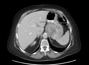
Abdominal CT scan demonstrating an 8.5 cm solid, well-marginated mass with non-homogeneous contrast enhancement along the fundus of the stomach extending to the gastroesophageal junction.
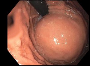
Endoscopic picture of intact gastric mucosa with bulging subepithelial mass in the cardia/fundus of the stomach visible on retroflexion view.
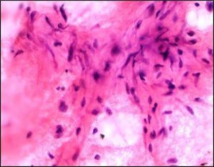
3A: FNA of submucosal lesion showing a few aggregates of cytologically bland spindle cells with myxoid background.
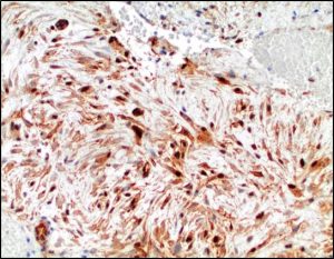
3B: Immunohistochemical stain for β-catenin shows strong and diffuse nuclear and cytoplasmic labeling in the tumor cells.
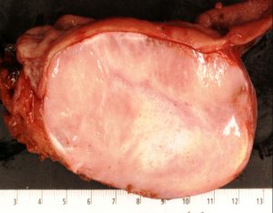
Cross section of the resected gastric mass reveals a well-demarcated intramural heterogenous mass with discrete contours. The overlying mucosa (upper portion of the picture) and the serosal surface (lower portion of the picture) are both intact. Esophageal-gastric junction is on the left side of the picture.
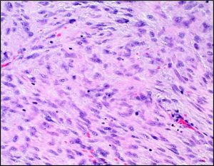
5A: High-power view of the central area of the lesion demonstrating a hypercellular storiform pattern of spindle cells with moderate nuclear pleomorphism and inconspicuous nucleoli.

Intermediate-power of hypocellular area from the periphery of the mass demonstrating spindle cells in a myxoid background containing dilated blood vessels with a staghorn pattern.
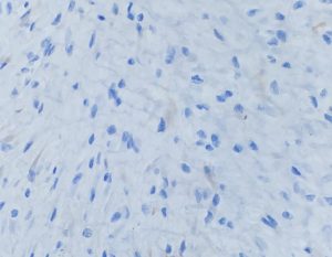
5C: Immunohistochemical stain for CD117 is negative.
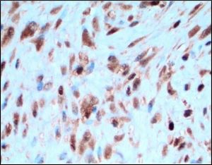
5D: Immunohistochemical stain for β-catenin shows cytoplasmic and strong nuclear labeling in the tumor cells.
Congratulations to the 2021 GIPS Abstract Award Winners
Winner: Chih-Ping Mao (Brigham and Women’s Hospital)
Integrated Clinicopathologic and Genomic Analysis to Predict Response to Neoadjuvant Treatment in Esophageal Cancer
First runner-up: Linyuan Wang (Emory University)
Analysis of KRAS Mutational Profiles in the gastrointestinal tract reveals site-specific alterations that can help distinguish pancreatobiliary and upper gastrointestinal primaries
Second runner-up: Christopher Bowman (University of California San Francisco)
Persistent or Recurrent Barrett’s Neoplasia After Endoscopic Therapy is Associated with DNA Content Abnormality Detected by DNA Flow Cytometric Analysis of Paraffin-Embedded Tissue